

MRI: Magnetic Resonance Imaging
Magnetic Resonance Imaging (MRI) generates images of the body without using radiation. Instead, an MRI uses a strong magnet and radio waves to create an image. There are no known harmful effects from the magnetic field or exposure to radio waves associated with creating MRI images.
Preparation
The presence of metal in or on your body may be a safety hazard for an MRI exam. There are also certain medical conditions that may prevent you from having an MRI exam. You will be asked a series of medical history questions to determine whether you may proceed with the exam. Please contact our staff to determine your eligibility for an MRI.
You will be asked to remove all metal or electronic objects from your body before the exam. These objects interfere with the magnetic field and can be very dangerous if taken into the exam room.
If you have ever had metal in your eyes you may need to have an X-ray of your head prior to undergoing an MRI.
Tell our staff at the time of your exam if you have any metal or electronic devices in or on your body including, but not limited to:
- Watches or jewelry
- Cell phones or PDAs
- Implanted electronic devices
- Implanted metallic joint prostheses, artificial heart valves, cochlear implants, hearing aids or metallic dentures.
Tell your technologist and your doctor if you are pregnant or suspect you may be pregnant. Your doctor may postpone the exam or choose an alternative exam.
No preparation is necessary prior to your exam.
Some patients may feel anxious due to the confining nature of the MRI scanner. If you feel this way, talk to your doctor. He or she may feel it necessary to prescribe a sedative prior to your exam to help you relax.
Your doctor or radiologist may request that your MRI scan be enhanced via the use of contrast material. If contrast is required, the technologist will start an intravenous (IV) line in your hand or arm to administer the contrast.
Although rare, there is a slight risk of an allergic reaction to contrast material. Therefore, you will be asked a series of questions about your medical history to determine whether you may receive contrast.
Most reactions are minor such as itchiness, hives, and nausea or vomiting. In very rare instances, an allergic reaction may cause swelling in your throat or other areas of your body. Tell your technologist or doctor immediately if you experience any of these symptoms during or after your exam.
Exam
The exam is painless and on average takes between 30 to 45 minutes, excluding preparation time. The amount of time varies depending on the type and number of exams for which you are scheduled.
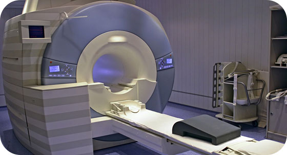
During the exam, you will be asked to lie on a movable table. A coil or small antenna-like device may be placed over the body part to be examined. Once you have been positioned, the technologist will move the table into the MRI scanner which is a tubular shaped machine.
The technologist monitors you throughout the procedure. A microphone system enables you to communicate with the technologist at all times. Upon request, you will also be given a device that will allow you to alert the technologist if you are having any difficulty during the procedure.
Results
When your exam is complete you may leave and resume regular activities. If a sedative is administered for your exam, you will need to arrange transportation home.
A radiologist will review your exam images and report the findings to your doctor within 24 hours. Your doctor will follow his/her office protocol to share the findings and next steps with you.